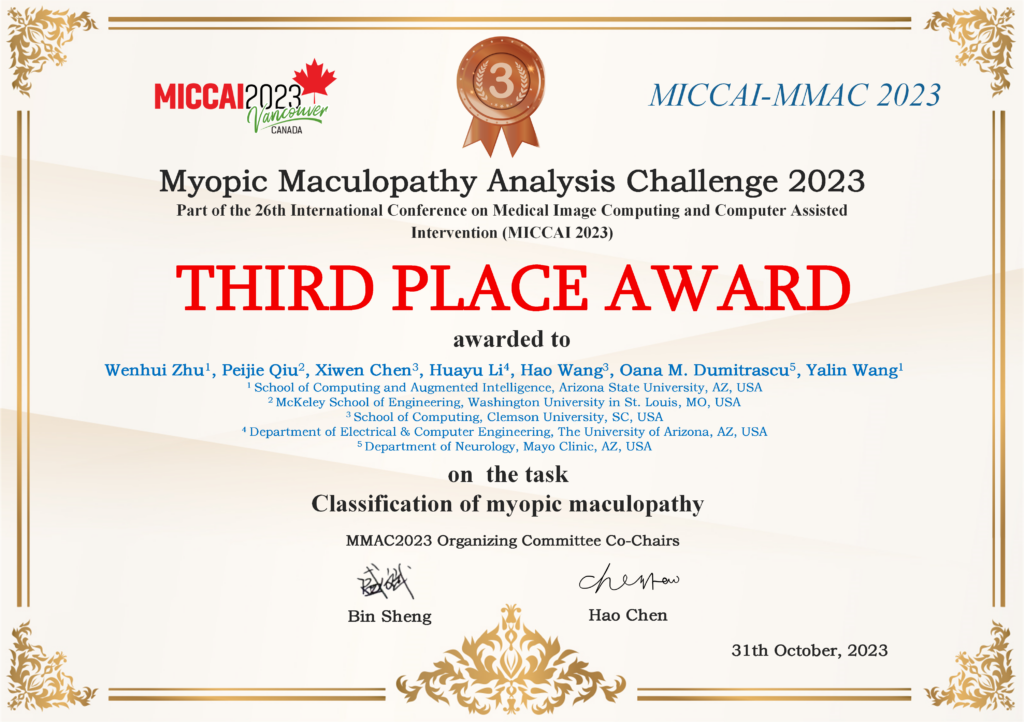
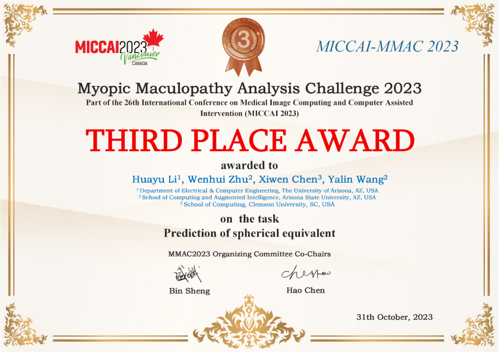
Wenhui won the 3rd in the MICCAI MMAC 2023 – Myopic Maculopathy Analysis Challenge in Both Task1 and 3.


Introduction
Myopia is a common oculopathy that affects large populations in the world. More seriously, myopia may further develop into high myopia in which the visual impairment mainly results from the development of different types of myopic maculopathy. In many countries, such as Japan, China, Denmark and the United States, myopic maculopathy is one of the leading causes of visual impairment and legal blindness worldwide. According to the severity, myopic maculopathy can be classified into five categories: no macular lesions, tessellated fundus, diffuse chorioretinal atrophy, patchy chorioretinal atrophy and macular atrophy. In addition, three additional “Plus” lesions are also defined and added to these categories: lacquer cracks (Lc), choroidal neovascularization (CNV), and Fuchs spot (Fs). Myopic maculopathy is likely to progress more quickly after the stage of tessellated fundus. It is estimated that about 90% of eyes with CNV showed a progression of myopic maculopathy. At present, fundus photography is a commonly used imaging modality in the diagnosis of myopic maculopathy. The advantage of fundus photography modality is that it can help doctors diagnose myopic maculopathy accurately and quickly, and can also achieve spherical equivalent prediction without mydriasis. Prompt screening and intervention are necessary to prevent the further progression of myopic maculopathy to avoid vision loss. However, the myopic maculopathy diagnosis is limited by the manual inspection process of image by image, which is time-consuming and relies heavily on the experience of ophthalmologists. Therefore, an effective computer-aided system is essential to help ophthalmologists analyze myopic maculopathy, further providing accurate diagnosis and reasonable intervention for this disease.
Aiming to advance the state-of-the-art in automatic myopic maculopathy analysis, we organize the myopic maculopathy analysis challenge. The challenge encourages researchers to develop algorithms for different tasks in myopic maculopathy analysis using fundus photography, including classification of myopic maculopathy, segmentation of myopic maculopathy plus lesions and spherical equivalent prediction. On the one hand, the classification and segmentation tasks for myopic maculopathy provide a basis for automated analysis of myopic maculopathy in clinical practice. On the other hand, higher degrees of myopia are associated with an increased risk of severe types of myopic maculopathy, so the prediction of spherical equivalent can help diagnose the risk of macular maculopathy. With this dataset, various algorithms can test their performance and make a fair comparison with other algorithms. As far as we know, it is the first dataset that covers the classification and segmentation of myopic maculopathy, and the prediction of spherical equivalent, with fundus images. We believe this challenge is an important milestone in myopic maculopathy analysis and hope that the challenge will drive forward innovation for automatic medical image analysis.
Reference link: https://codalab.lisn.upsaclay.fr/competitions/12441 (task 1)
Reference link: https://codalab.lisn.upsaclay.fr/competitions/12477 (task 3)