Hippocampal and Ventricular Differences in 804 ADNI subjects mapped with Multivariate Tensor-Based Morphometry
Yalin Wang, Yang Song, Yi-Yu Chou, Arthur W. Toga, Paul M. Thompson and the Alzheimer’s Disease Neuroimaging Initiative (ADNI)
Abstract
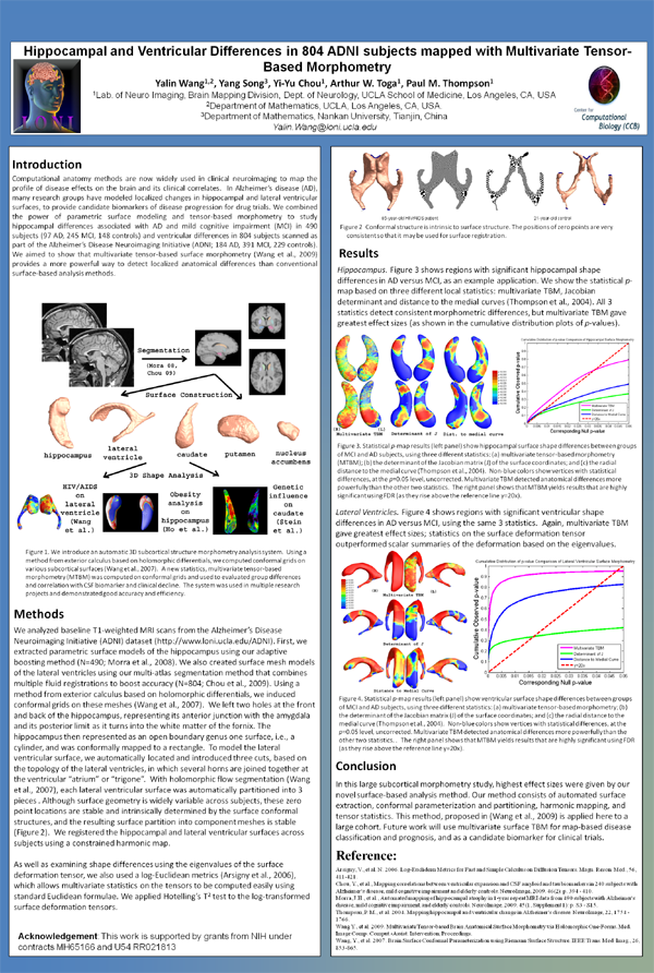
Computational anatomy methods are now widely used in clinical neuroimaging to map the profile of disease effects on the brain and its clinical correlates. In Alzheimer’s disease (AD), many research groups have modeled localized changes in hippocampal and lateral ventricular surfaces, to provide candidate biomarkers of disease progression for drug trials. We combined the power of parametric surface modeling and tensor-based morphometry to study hippocampal differences associated with AD and mild cognitive impairment (MCI) in 490 subjects (97 AD, 245 MCI, 148 controls) and ventricular differences in 804 subjects scanned as part of the Alzheimer’s Disease Neuroimaging Initiative (ADNI; 184 AD, 391 MCI, 229 controls). We aimed to show that multivariate tensor-based surface morphometry (Wang et al., 2009) provides a more powerful way to detect localized anatomical differences than conventional surface-based analysis methods.
Figures (click on each for a larger version):
Related Publications
- Wang Y, Song Y, Chou Y, Toga AW, Thompson, PM, “Hippocampal and Ventricular Differences in 804 ADNI subjects mapped with Multivariate Tensor-Based Morphometry”, 16th Annual Meeting of the Organization for Human Brain Mapping. 2010: Barcelona, Spain. (This paper won a planetary talk)