Automated Retinal Imaging Analysis for Alzheimers Disease Screening
Dumitrascu OM, Zhu W, Qiu P, Nandakumar K, Wang Y
Abstract
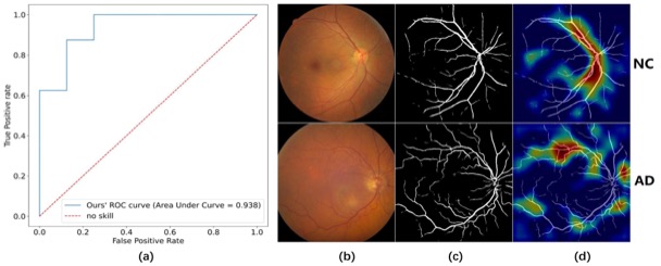
Alzheimer’s Disease (AD), the most common cause of de- mentia, has increasing prevalence with vast societal and pub- lic health implications. There is a critical unmet need to develop biomarkers for early AD diagnosis. Recent scientific advances underscore retinal vascular changes and retinal ab- normal protein deposition mirroring the changes in the AD brain. We have previously shown that retinal vascular tortu- osity and inflection correlate with neurocognitive dysfunction and may predict AD. Retinal fundus photography is a cost-effective and high-resolution imaging tool to study reti- nal vascular changes in AD and emerge as a non-invasive biomarker for early AD diagnosis and monitoring. Hand- crafted identification of the retinal vascular features on color fundus images is laborious, subjective, and prone to bias. Hence, developing automated retinal imaging tools has at- tracted strong research interest. Here, we leverage deep neural networks to develop an automatic framework to clas- sify AD and extract AD retinal fundus imaging biomarkers using weakly supervised localization and Gradient-weighted Class Activation Mapping.
Figures (click on each for a larger version):