Enhancing Alzheimer’s Diagnosis: Leveraging Anatomical Landmarks in Graph Convolutional Neural Networks on Tetrahedral Meshes
Chen Y, Farazi M, Yang Z, Fan Y, Ashton N, Reiman E, Su Y, Wang Y
Abstract
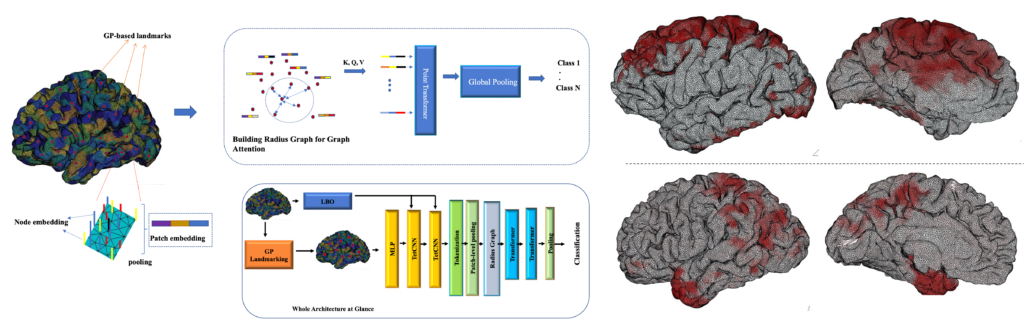
Alzheimer’s disease (AD) is a major neurodegenerative condition that affects millions around the world. As one of the main biomarkers in the AD diagnosis procedure, brain amyloid positivity is typically identified by positron emission tomography (PET), which is costly and invasive. Brain structural magnetic resonance imaging (sMRI) may provide a safer and more convenient solution for the AD diagnosis. Recent advances in geometric deep learning have facilitated sMRI analysis and early diagnosis of AD. However, determining AD pathology, such as brain amyloid deposition, in preclinical stage remains challenging, as less significant morphological changes can be observed. As a result, few AD classification models are generalizable to the brain amyloid positivity classification task. Blood-based biomarkers (BBBMs), on the other hand, have recently achieved remarkable success in predicting brain amyloid positivity and identifying individuals with high risk of being brain amyloid positive. However, individuals in medium risk group still require gold standard tests such as Amyloid PET for further evaluation. Inspired by the recent success of transformer architectures, we propose a geometric deep learning model based on transformer that is both scalable and robust to variations in input volumetric mesh size. Our work introduced a novel tokenization scheme for tetrahedral meshes, incorporating anatomical landmarks generated by a pre-trained Gaussian process model. Our model achieved superior classification performance in AD classification task. In addition, we showed that the model was also generalizable to the brain amyloid positivity prediction with individuals in the medium risk class, where BM alone cannot achieve a clear classification. Our work may enrich geometric deep learning research and improve AD diagnosis accuracy without using expensive and invasive PET scans.
Figures (click on each for a larger version):