Applying Surface-Based Morphometry to Study Ventricular Abnormalities of Cognitively Unimpaired Subjects Prior to Clinically Significant Memory Decline
Dong Q*, Zhang W*, Stonnington CM, Wu J, Gutman BA, Chen K, Su Y, Baxter LC, Thompson PM, Reiman EM, Caselli RJ, Wang Y, ADNI Group
Abstract
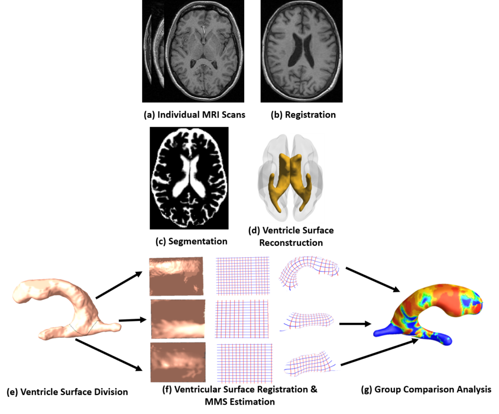
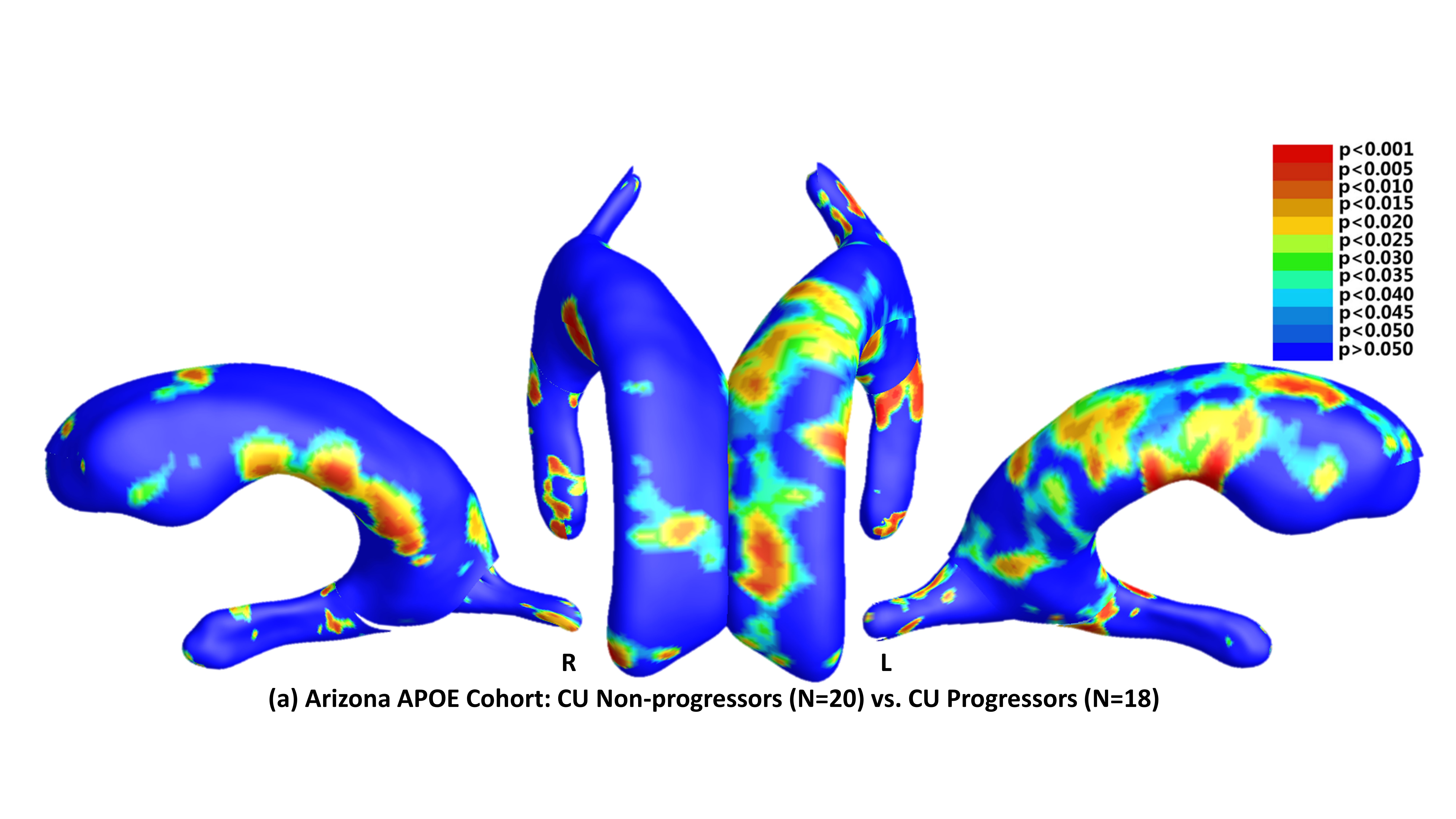
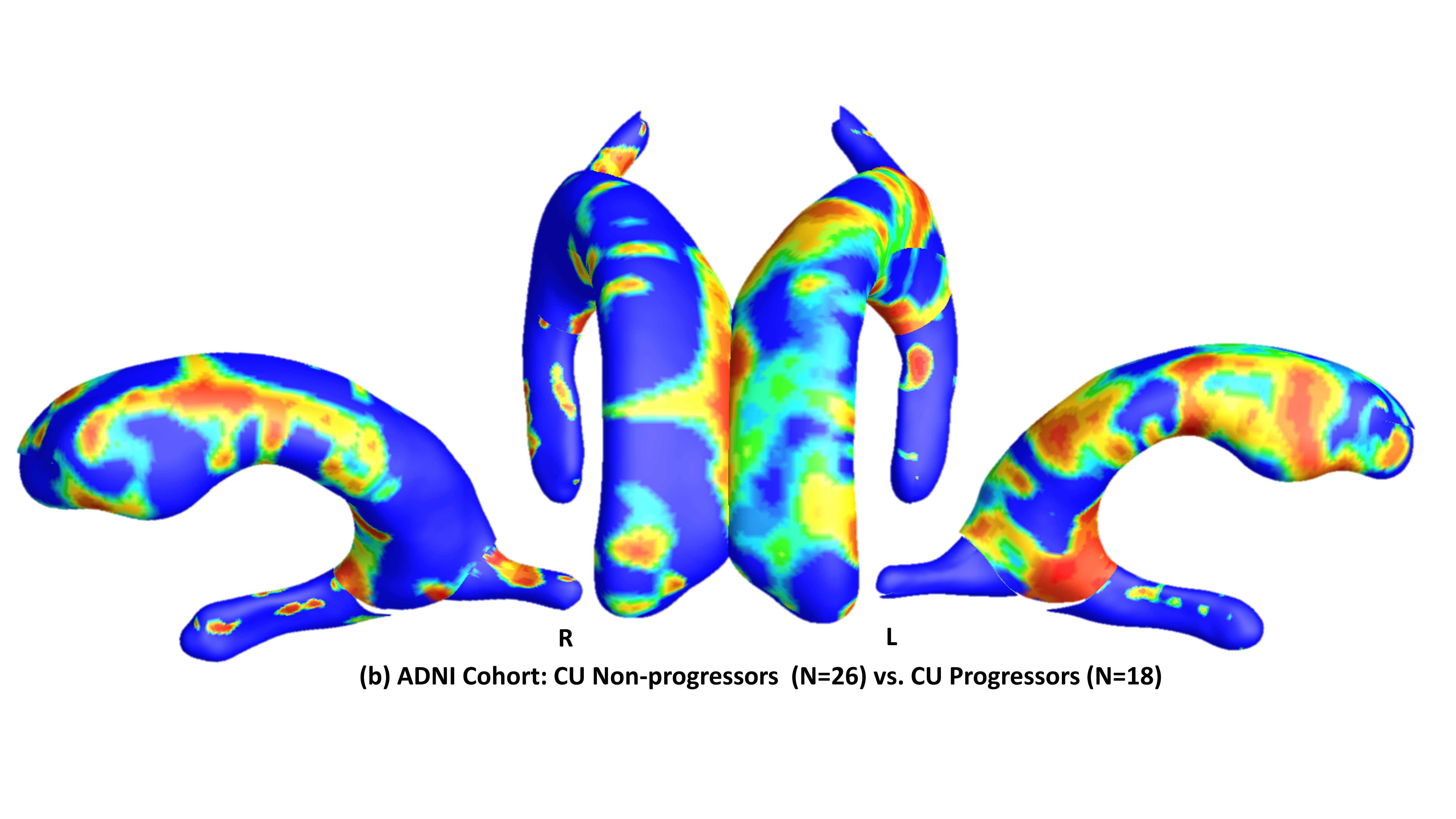
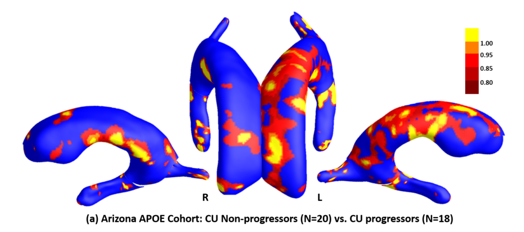
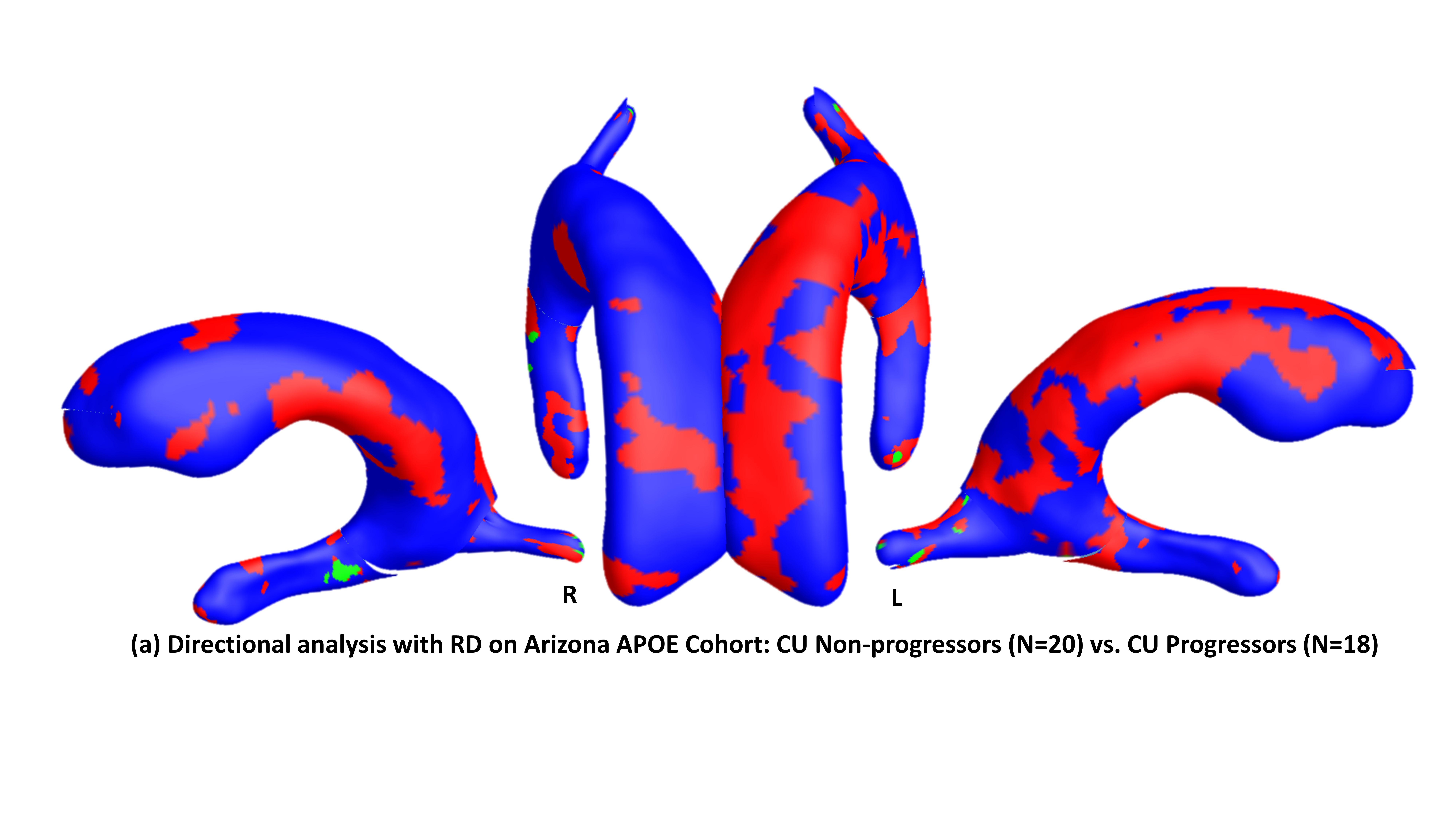
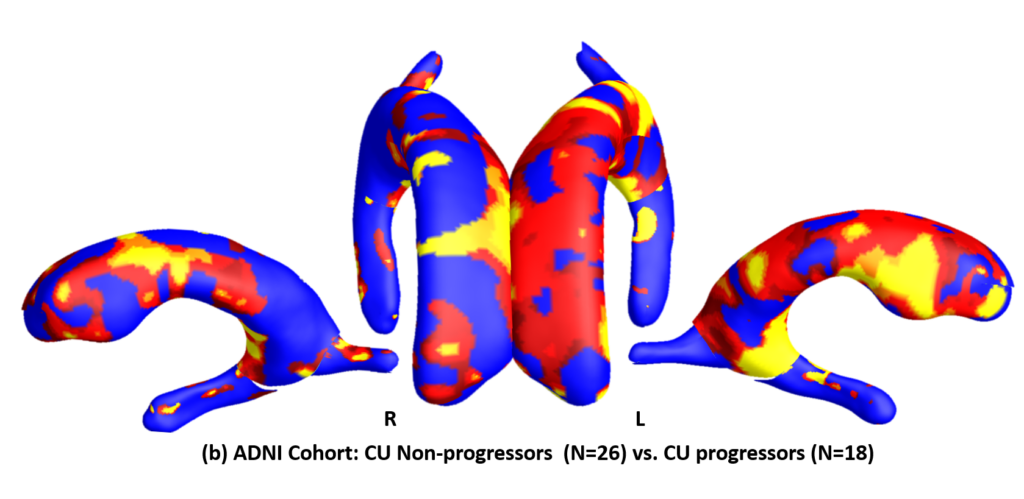
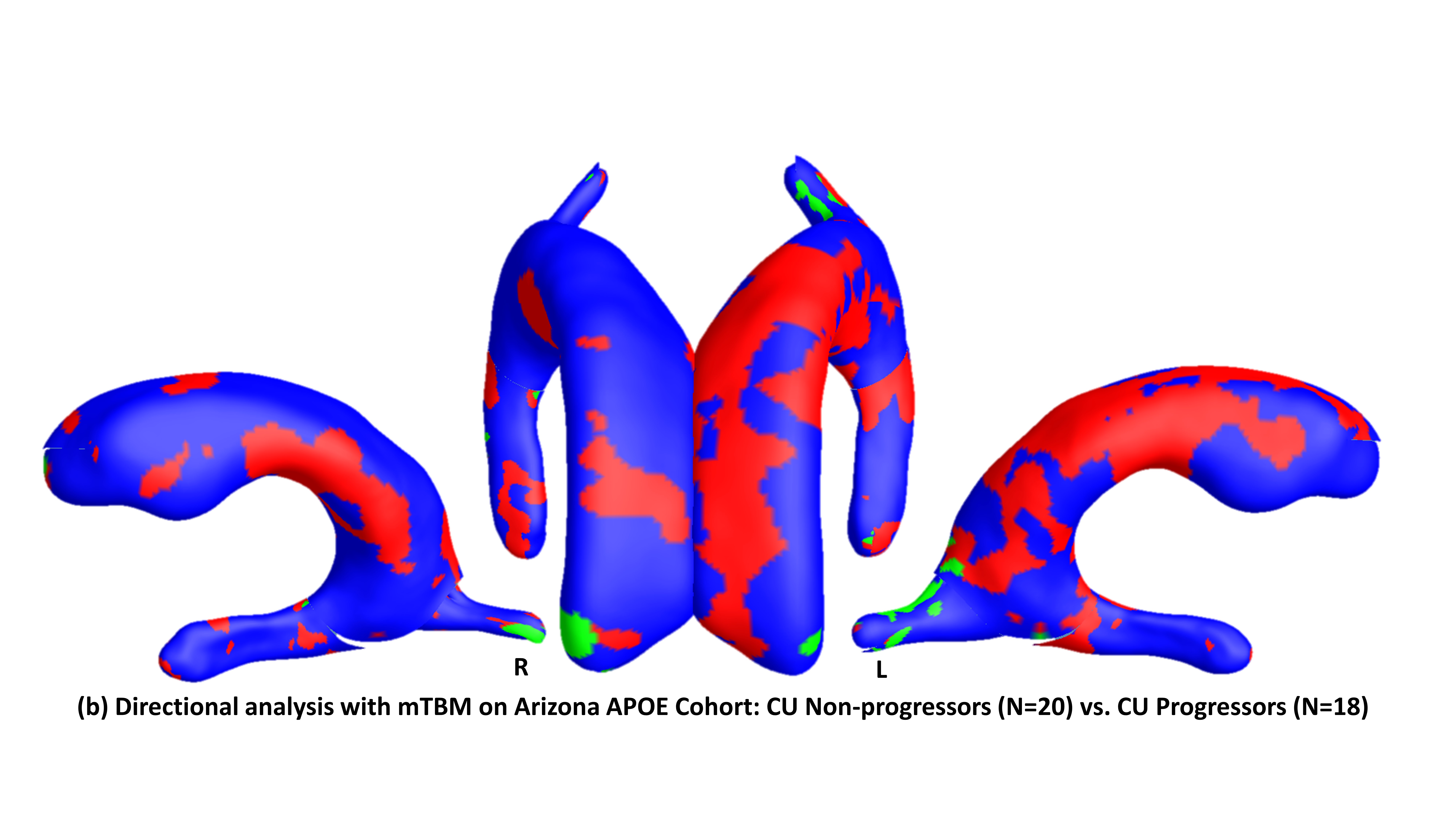
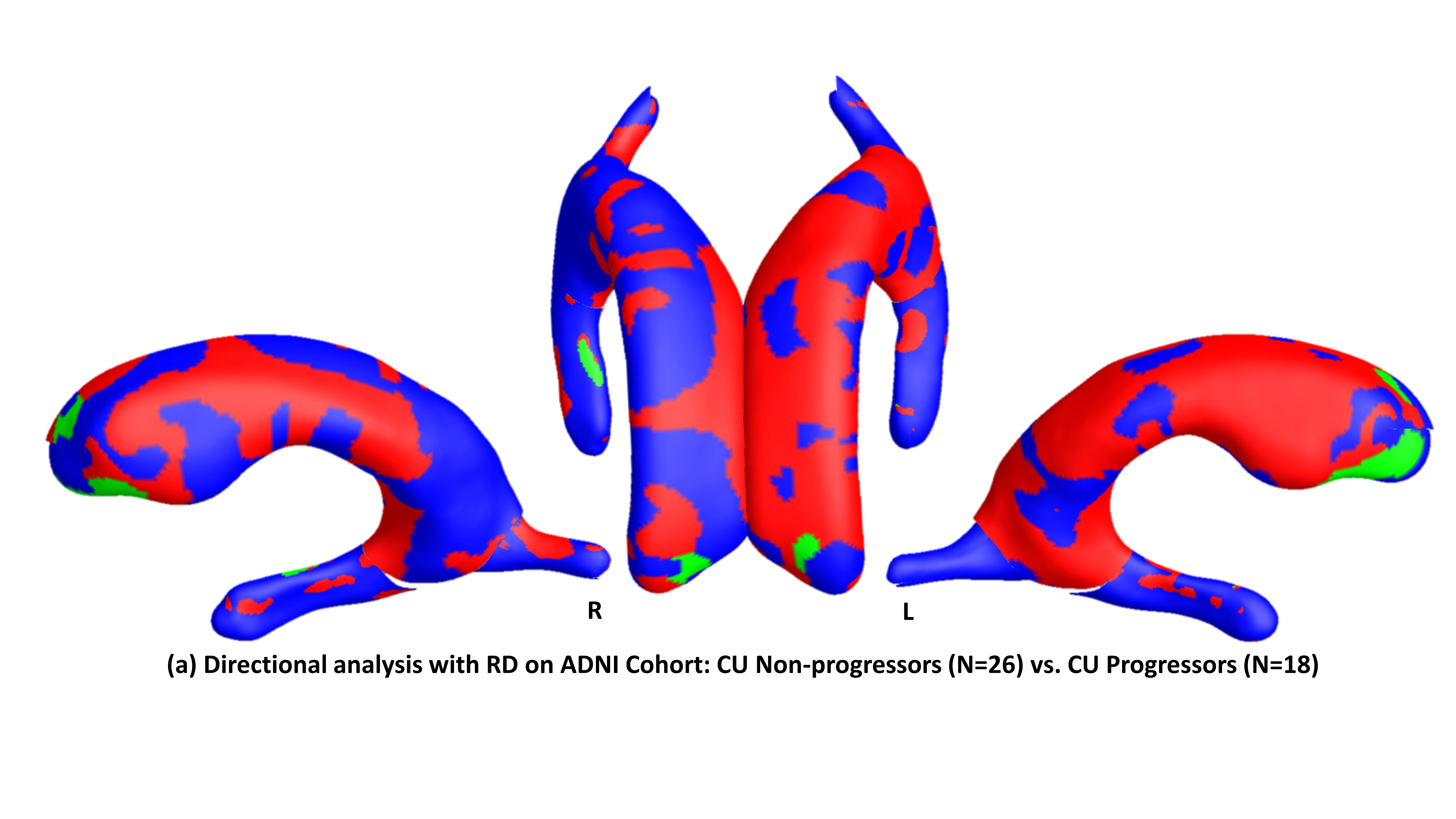
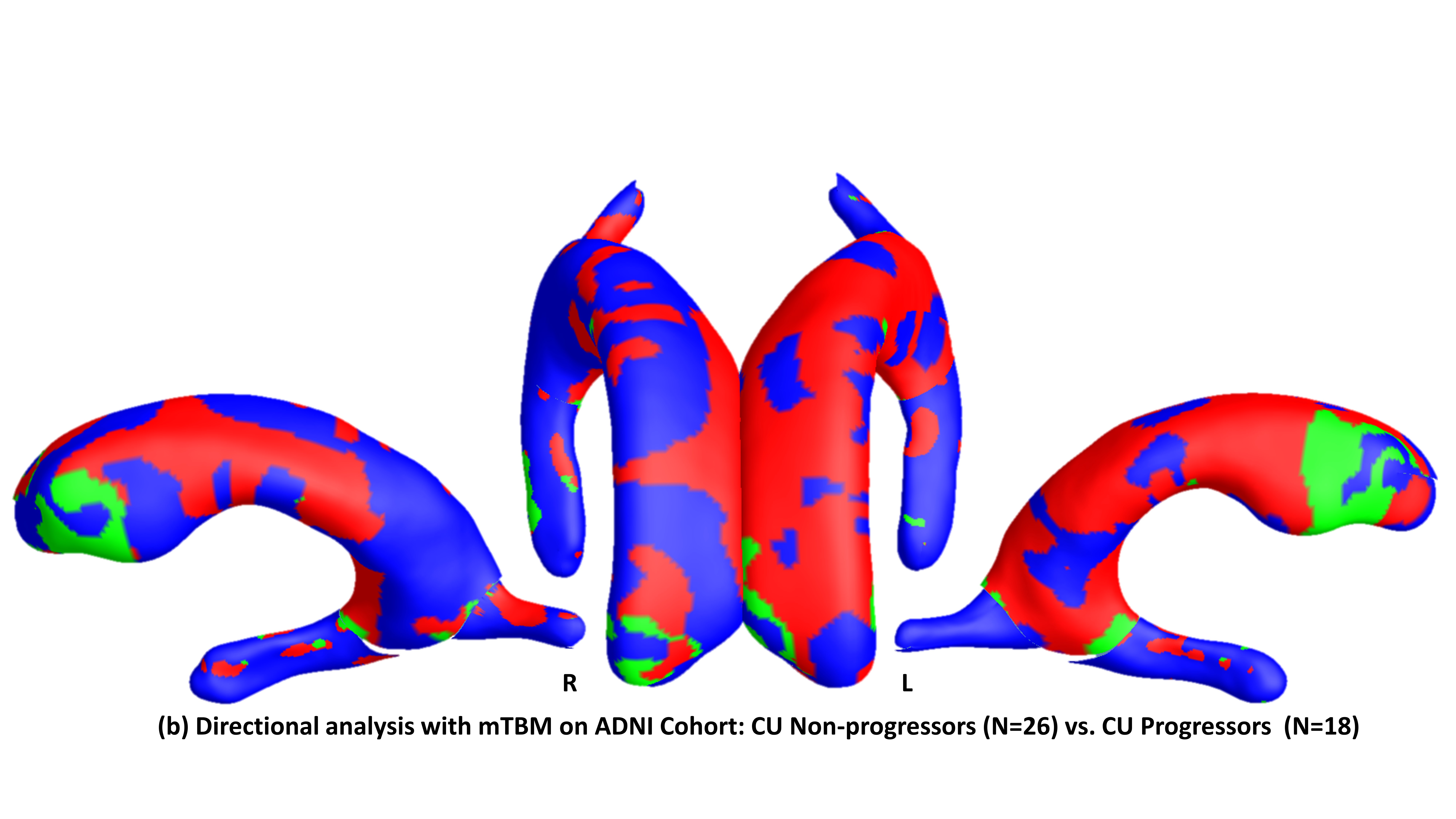
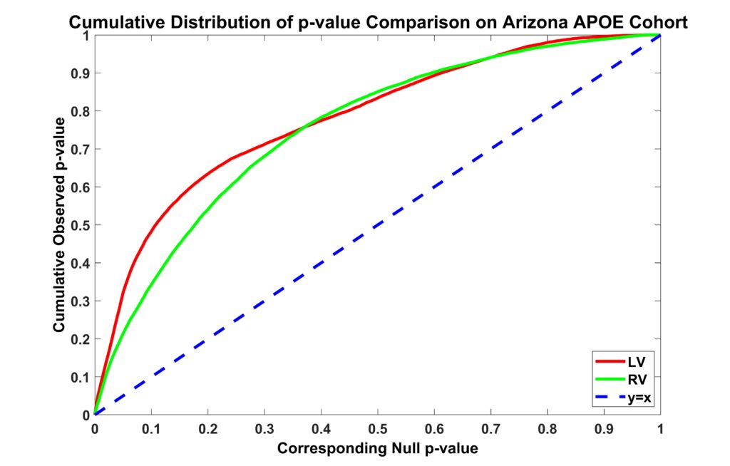
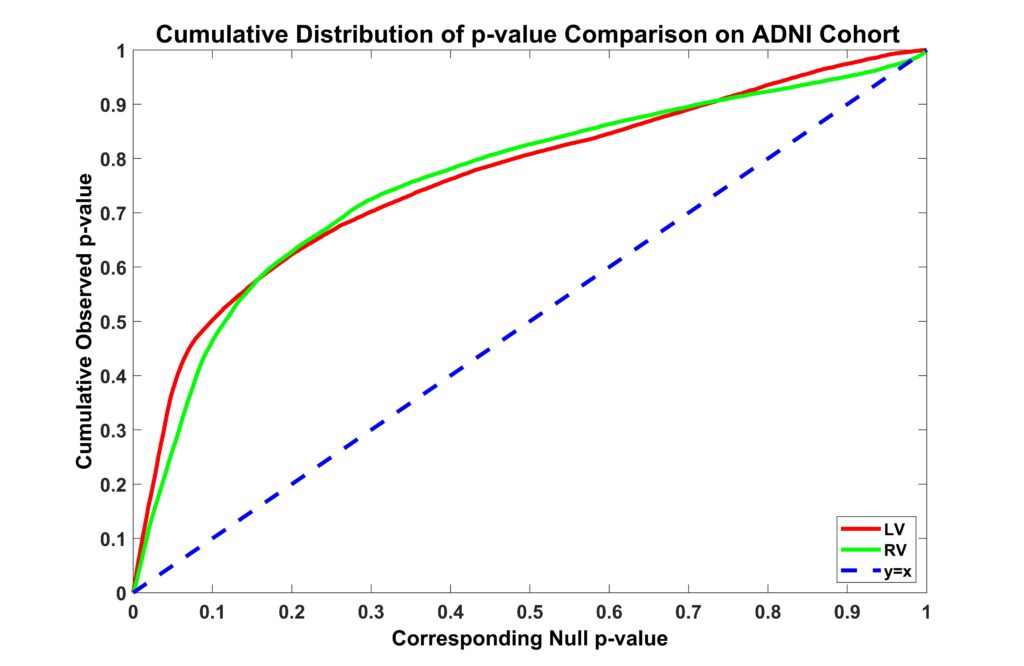
Ventricular volume (VV) is a widely used structural magnetic resonance imaging (MRI) biomarker in Alzheimer’s disease (AD) research. Abnormal enlargements of VV can be detected before clinically significant memory decline. However, VV does not pinpoint the details of subregional ventricular expansions. Here we introduce a ventricular morphometry analysis system (VMAS) that generates a whole connected 3D ventricular shape model and encodes a great deal of ventricular surface deformation information that is inaccessible by VV. VMAS contains an automated segmentation approach and surface-based multivariate morphometry statistics. We applied VMAS to two independent datasets of cognitively unimpaired (CU) groups. To our knowledge, it is the first work to detect ventricular abnormalities that distinguish normal aging subjects from those who imminently progress to clinically significant memory decline. Significant bilateral ventricular morphometric differences were first shown in 38 members of the Arizona APOE cohort, which included 18 CU participants subsequently progressing to the clinically significant memory decline within 2 years after baseline visits (progressors), and 20 matched CU participants with at least 4 years of post-baseline cognitive stability (non-progressors). VMAS also detected significant differences in bilateral ventricular morphometry in 44 Alzheimer’s disease Neuroimaging Initiative (ADNI) subjects (18 CU progressors vs. 26 CU non-progressors) with the same inclusion criterion. Experimental results demonstrated that the ventricular frontal horn regions were affected bilaterally in CU progressors, and more so on the left. VMAS may track disease progression at subregional levels and measure the effects of pharmacological intervention at a preclinical stage.
Figures (click on each for a larger version):
Related Publications
- Dong Q*, Zhang W*, Wu J, Li B, Schron EH, McMahon T, Shi J, Gutman BA, Chen K, Baxter LC, Thompson PM, Reiman EM, Caselli RJ, Wang Y, “Applying Surface-Based Hippocampal Morphometry to Study APOE-e4 Allele Dose Effects in Cognitively Unimpaired Subjects”, NeuroImage: Clinical, 2019, 22:101744
